Feasibility of Different Tumor Delineation Approaches for 18F-PSMA-1007 PET/CT Imaging in Prostate Cancer Patients.
Mittlmeier LM, Brendel M, Beyer L, et al.
Delineation of PSMA-positive tumor volume on PET using PSMA-ligands is of highest clinical interest as changes of PSMA-PET/CT-derived whole tumor volume (WTV) have shown to correlate with treatment response in metastatic prostate cancer patients.
Read complete articleSo far, WTV estimation was performed on PET using 68Ga-labeled ligands; nonetheless, 18F-labeled PET ligands are gaining increasing importance due to advantages over 68Ga-labeled compounds. However, standardized tumor delineation methods for 18F-labeled PET ligands have not been established so far. As correlation of PET-based information and morphological extent in osseous and visceral metastases is hampered by morphological delineation, low contrast in liver tissue and movement artefacts, we correlated CT-based volume of lymph node metastases (LNM) and different PET-based delineation approaches for thresholding on 18F-PSMA-1007 PET.
Methods
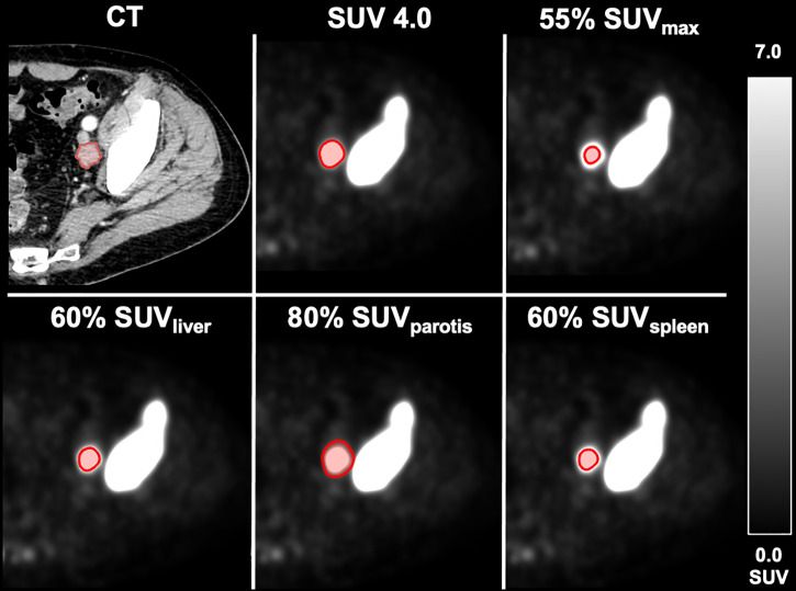
Fifty patients with metastatic prostate cancer, 18F-PSMA-1007 PET/CT and non-bulky LNM (short-axis diameter ≥10mm) were included. Fifty LNM were volumetrically assessed on contrast-enhanced CT (volumetric reference standard). Different approaches for tumor volume delineation were applied and correlated with the reference standard: I) fixed SUV threshold, II) isocontour thresholding relative to SUVmax (SUV%), and thresholds relative to III) liver (SUVliver), IV) parotis (SUVparotis) and V) spleen (SUVspleen).
Results
A fixed SUV of 4.0 (r=0.807, r2 = 0.651, p<0.001) showed the best overall association with the volumetric reference. 55% SUVmax (r=0.627, r2 = 0.393, p<0.001) showed highest association using an isocontour-based threshold. Best background-based approaches were 60% SUVliver (r=0.715, r2 = 0.511, p<0.001), 80% SUVparotis (r=0.762, r2 = 0.581, p<0.001) and 60% SUVspleen (r=0.645, r2 = 0.416, p<0.001). Background tissues SUVliver, SUVparotis & SUVspleen did not correlate (p>0.05 each). Recently reported cut-offs for intraprostatic tumor delineation (isocontour 44% SUVmax, 42% SUVmax and 20% SUVmax) revealed inferior association for LNM delineation.
Conclusions
A threshold of SUV 4.0 for tumor delineation showed highest association with volumetric reference standard irrespective of potential changes in PSMA-avidity of background tissues (e. g. parotis). This approach is easily applicable in clinical routine without specific software requirements. Further studies applying this approach for total tumor volume delineation are initiated.
Front Oncol. 2021;11:663631. Published 2021 May 21