DPD Quantification in Cardiac Amyloidosis: A Novel Imaging Biomarker.
Scully PR, Morris E, Patel KP, Treibel TA, Burniston M, Klotz E, Newton JD, Sabharwal N, Kelion A, Manisty C, Kennon S, Ozkor M, Mullen M, Hartman N, Elliott PM, Pugliese F, Hawkins PN, Moon JC, Menezes LJ.
Transthyretin-related cardiac amyloidosis is common and can be diagnosed noninvasively using bone scintigraphy; interpretation, however, relies on planar images. SPECT/CT imaging offers 3-dimensional visualization.
Read complete articleObjectives
To assess whether single-photon emission computed tomography (SPECT/CT) quantification of bone scintigraphy would improve diagnostic accuracy and offer a means of quantifying amyloid burden.
Methods
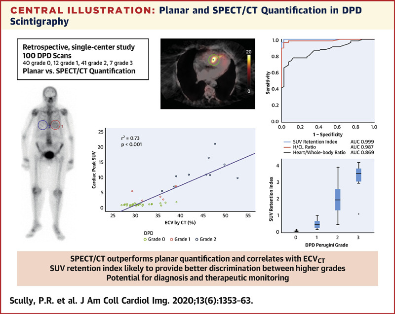
This was a single-center, retrospective analysis of 99mTc-3,3-diphosphono-1,2-propanodicarboxylic acid (DPD) scans reported using the Perugini grading system (0 = negative; 1 to 3 = increasingly positive). Conventional planar quantification techniques (heart/contralateral lung, and heart/whole-body retention ratios) were performed. Heart, adjacent vertebra, paraspinal muscle and liver peak standardized uptake values (SUVpeak) were recorded from SPECT/CT acquisitions. An SUV retention index was also calculated: (cardiac SUVpeak/vertebral SUVpeak) × paraspinal muscle SUVpeak. In a subgroup of patients, SPECT/CT quantification was compared with myocardial extracellular volume quantification by CT imaging (ECVCT).
Results
A total of 100 DPD scans were analyzed (patient age 84 ± 9 years; 52% male): 40 were Perugini grade 0, 12 were grade 1, 41 were grade 2, and 7 were grade 3. Cardiac SUVpeak increased from grade 0 to grade 2; however, it plateaued between grades 2 and 3 (p < 0.001). Paraspinal muscle SUVpeak increased with grade (p < 0.001), whereas vertebral SUVpeak decreased (p < 0.001). The composite parameter of SUV retention index overcame the plateauing of the cardiac SUVpeak and increased across all grades (p < 0.001). Cardiac SUVpeak correlated well (r2 = 0.73; p < 0.001) with ECVCT. Both the cardiac SUVpeak and SUV retention index had excellent diagnostic accuracy (area under the curve [AUC]: 0.999). The heart to contralateral lung ratio performed the best of the planar quantification techniques (AUC: 0.987).
Conclusions
SPECT/CT quantification in DPD scintigraphy is possible and outperforms planar quantification techniques. Differentiation of Perugini grade 2 or 3 is confounded by soft tissue uptake, which can be overcome by a composite SUV retention index. This index can help in the diagnosis of cardiac amyloidosis and may offer a means of monitoring response to therapy.
Keywords
DPD scintigraphy; SPECT/CT quantification; cardiac amyloidosis.
Copyright © 2020 The Authors. Published by Elsevier Inc. All rights reserved.
JACC Cardiovasc Imaging. 2020 Jun;13(6):1353-1363. doi: 10.1016/j.jcmg.2020.03.020. Erratum in: JACC Cardiovasc Imaging. 2021 Jan;14(1):318-319.