Comparison of standardized uptake values between 99mTc-HDP SPECT/CT and 18F-NaF PET/CT in bone metastases of breast and prostate cancer
Samuli Arvola et al.
Methods
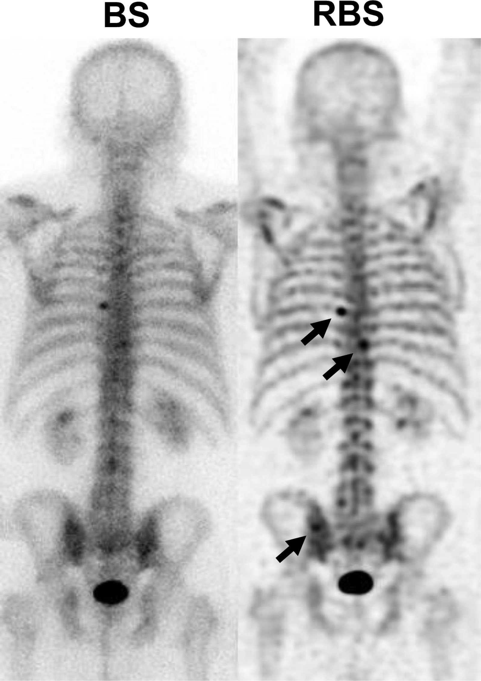
Twenty-six breast and 27 prostate cancer patients at high risk of bone metastases underwent both 99mTc-HDP SPECT/CT and 18F-NaF PET/CT within 14 days of each other. The SPECT and PET data were reconstructed using ordered-subset expectation-maximization algorithms achieving quantitative images. Metastatic and benign skeletal lesions visible in both data sets were identified, and their maximum, peak, and mean SUVs (SUVmax, SUVpeak, and SUVmean) were determined. SUV ratios (SUVRs) between the lesions and adjacent normal appearing bone were also calculated. Linear regression was used to evaluate the correlations between the SUVs of SPECT and PET and Bland-Altman plots to evaluate the differences between the SUVs and SUVRs of SPECT and PET.
Results
A total of 231 skeletal lesions, 129 metastatic and 102 benign, were analyzed. All three SUV measures correlated very strongly between SPECT and PET (R2 ≥ 0.80, p < 0.001) when all lesions were included, and the PET SUVs were significantly higher than SPECT SUVs (p < 0.001). The median differences were 21%, 12%, and 19% for SUVmax, SUVpeak, and SUVmean, respectively. On the other hand, the SUVRs were similar between SPECT and PET with median differences of 2%, − 9%, and 2% for SUVRmax, SUVRpeak, and SUVRmean, respectively.
Conclusion
The strong correlation between SUVs and similar SUVRs of 99mTc-HDP SPECT/CT and 18F-NaF PET/CT demonstrate that SPECT is an applicable tool for clinical quantification of bone metabolism in osseous metastases in breast and prostate cancer patients.