Hermia | PET/CT, SPECT/CT, and PET/MR
The Hermia Software Suite offers a high-performance and highly intuitive vendor-neutral software for the display and analysis of PET/CT, SPECT/CT, and PET/MR.

Supporting fast and accurate reviewing
Hermia provides a powerful and intuitive toolkit for the display and analysis of nuclear medicine images. Supported data includes PET/CT, SPECT/CT, PET/MR, RTDOSE and 2D nuclear medicine images.
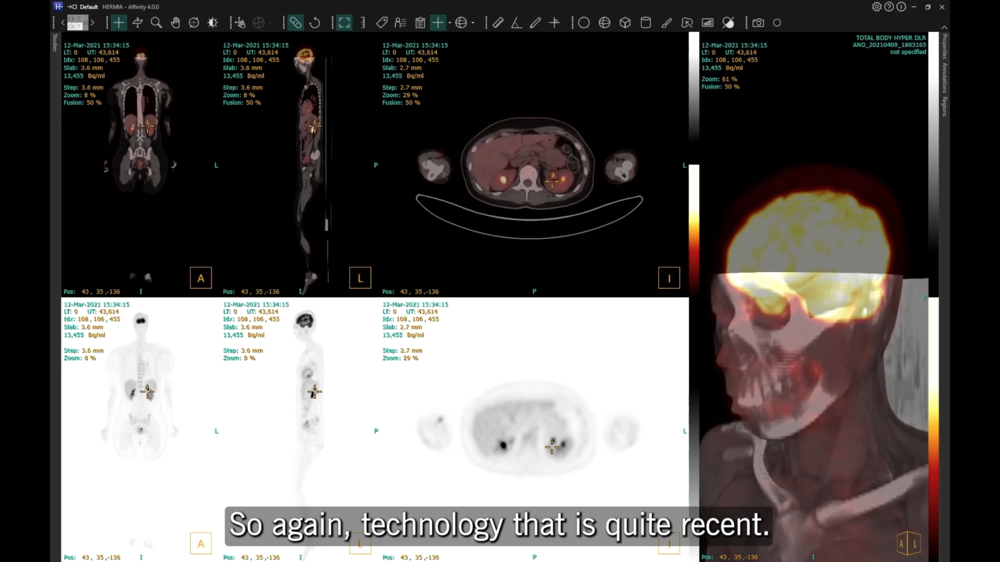
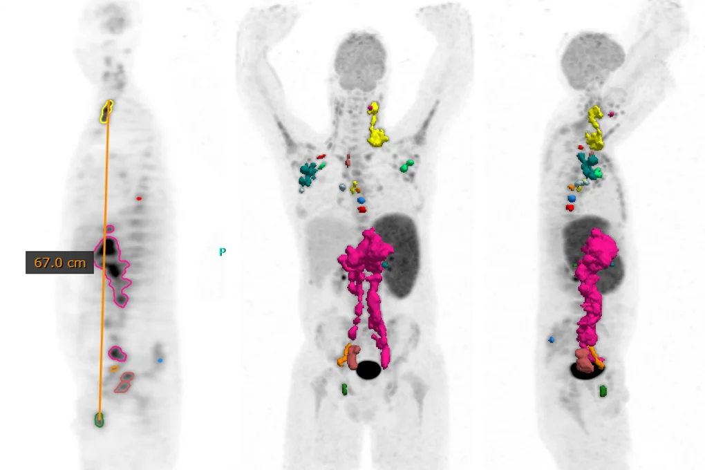
 Data from a Siemens Biograph Vision Quadra Whole-Body PET/CT provided by Inselspital Bern, Switzerland
-
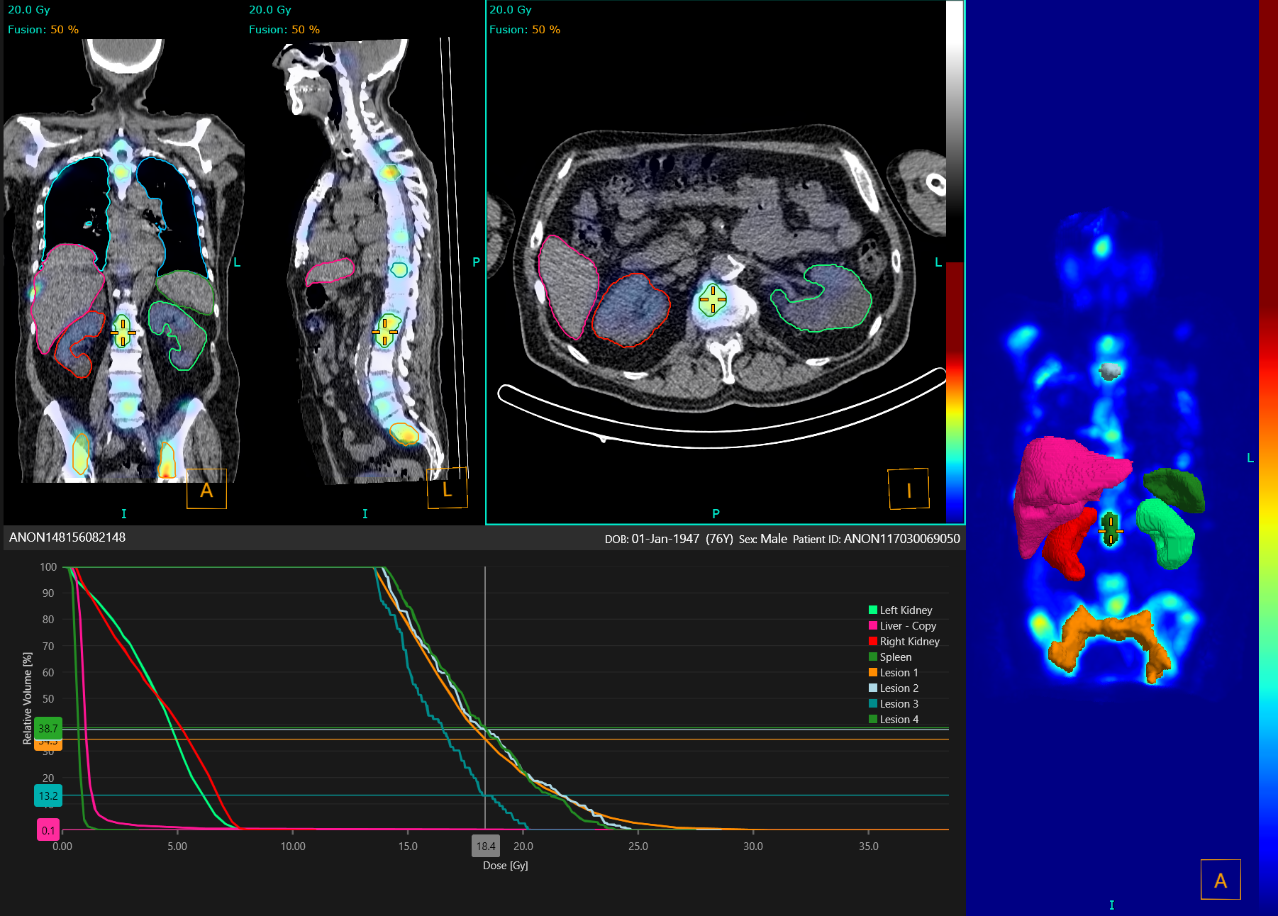
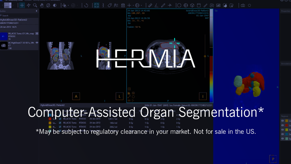
Computer-assisted* organ segmentation
Liver, kidney, spleen, and lung segmentation algorithm runs in the background while you work, saving time during dosimetry analysis and reducing operator variability.
-
Personalized user experience
Completely customizable workflows and layouts to suit your reporting needs.
-
High throughput reporting
After selecting patient images at study level, Hermia will pick the best images to display and choose the most appropriate workflow based on your requirements.
-
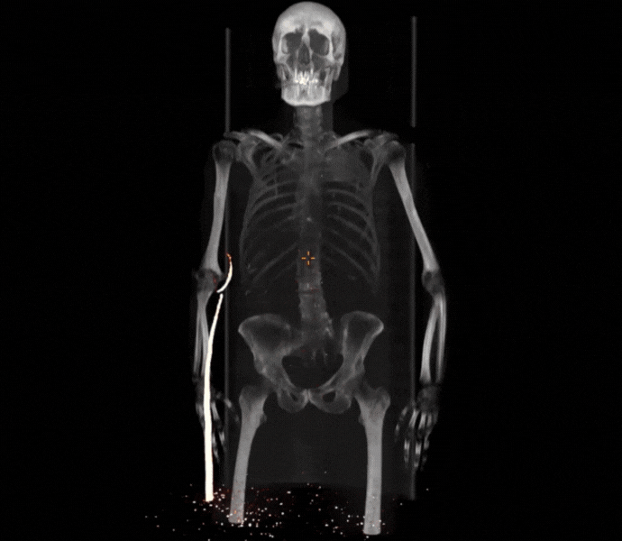
Multimodality visualization
Load images from multiple time points and modalities in any combination. Transparency alpha blending gives clearer visualization in fusion to maximise image information.
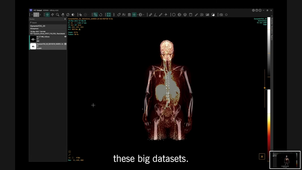
Dynamic PET/CT data from the Siemens Quadra in Hermia software
Whole-body PET-CT data from United Imaging's scanner uExplorer in Hermia software
Computer-assisted organ segmentation
Liver, kidney, spleen, and lung segmentation algorithm runs in the background while you work, saving time during dosimetry analysis and reducing operator variability. All segmentation can be adjusted by the user. Assess region dose coverage using dose-volume histograms and export regions as DICOM SEG, RTSS, or NIfTI file format.
High throughput reporting
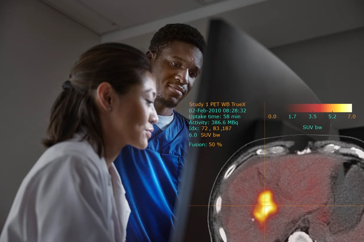
Hermia exploits the latest computing technology to accelerate image review. Select patient images at study level; Hermia will pick the best images to display and choose the most appropriate workflow based on your requirements. SUV mouse mode for quick and easy SUVmax localization. Sub-second automatic registration gives easy comparison
between multiple time points.
Advanced oncology tools
Quantify total metabolic tumor volume even in the most challenging cases with a versatile toolkit including single click lesion segmentation, thresholding, and splitting tools. Use the blob-splitter to help differentiate clinically relevant uptake from physiological or edit with a 3D paintbrush. Measure ‘DMax’* for disseminated disease to quantify spread. Flexible and intuitive, region drawing has never been so easy.
Local registration
Refine image alignment by focusing on a specific area of interest. Automatic registration is redone using only the image information surrounding the triangulation point, allowing precise comparison over multiple time points in breathing motion affected areas.
Personalized user experience
Completely customizable workflows and layouts to suit your reporting needs. User preferences including mouse and keyboard shortcuts, crosshair options, and SUV statistics will follow your profile no matter where you log in. Specify the information to be displayed alongside images including uptake time*, administered activity, voxel index coordinates, and more.
Multimodality visualization
Load images from multiple time points and modalities in any combination. Align and fuse as many layers as needed for the best display. Transparency alpha blending gives clearer visualization in fusion to maximise image information. Reorient images in all planes with live graphical updates for fast 3D manipulation.
*May be subject to regulatory clearance in your market. Not for sale in the USA.
Schedule a free consultation
To support all your clinical workflows
Browse to read more about all the many possibilities offered by Hermia– our ALL-IN-ONE state-of-the-art software suite. You can pick and choose the specialities and tools adapted to your current clinical needs and scale up whenever new possibilities arise.
-
Read more
SPECT reconstruction
Hermia's SPECT reconstruction is optimized for speed and a wide range of procedures, radio-pharmaceuticals and collimators, making it possible to improve image quality while reducing dose and acquisition time from all your SPECT/CT cameras.
-
Read more
Oncology
Handle all your oncology work from diagnostic imaging, reporting to therapy for all modalities and scanners.
-
Read more
Theranostics
Theranostics is a very promising personalized approach to treating cancer, using diagnostic imaging to identify if target receptors are present on cancer cells, followed by precision internal radiation treatment that targets these receptors.
-
Read more
Dosimetry
Dosimetry is becoming more important as nuclear medicine is moving towards therapy. Regardless of where your practice is at, we have got you covered with the tools you need to easily plan personalized radionuclide treatment for your patients.
-
Read more
Neurology
Hermia Neurology has a fully automated 'single click' workflow. Data is processed, quantified and displayed within seconds with minimal user intervention.
-
Read more
Pneumology
Anatomically accurate lung function and volumes with Lung Lobe Quantification and fast and easy Lung SPECT VQ image display and analysis
-
Read more
Cardiology
State-of-the-art product line for cardiology, including third-party software with Invia Corridor4DM and Cedars-Sinai Cardiac Suite.
-
Read more
IT Solutions
Our Software and Hardware solutions have been designed to integrate seamlessly to your current workflow and systems and to offer the possibility to grow with your organization. The result is an improved user experience where the systems work for the user and not the other way around.