Hermes Medical Solutions releases a new version of the Hermia Multimodality Viewer software in North America
Hermes Medical Solutions (HMS), global market leader in molecular imaging and dosimetry software solutions, launches the latest version of its Hermia Multimodality Viewing Software in Canada and in the USA.
Please accept marketing cookies to see this video content.
“This new release gives North American users a new level of customization and personalization of settings and workflow for PET-CT, PET-MR and SPECT-CT reviewing, in a modern and intuitive software platform. It’s another step towards our goal of providing molecular imaging and nuclear medicine departments with user-friendly software that’s compatible with scanners from any manufacturer and enables staff to work efficiently and flexibly.” Tom Francke, CEO of Hermes Medical Solutions.
Discover below some of the highlights of the latest version of the Hermia Multimodality Viewer software, which has just been released in Canada and in the US.
A new world of customization for your working preferences
Hermia makes it quick and easy to carry out routine Viewing, as well as more complex comparative analysis, thanks to fully configurable user-defined work- flows and screen layouts.
This new release of the Hermia Multimodality Viewer brings many new shortcut options for keyboard and mouse to simplify and speed up your work. Your personal shortcuts list is easy to access and update.
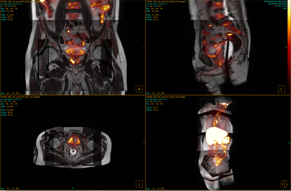
Advanced visualization with a true 3D multimodality viewer
Featuring our unique Deep Fusion TechnologyTM, Hermia Multimodality Viewer is designed to handle multiple time points, bed positions and modalities with ease. It allows any number of fused image layers to support expert reading – bringing new insights for confident diagnosis. 3D data from any modality, and in any combination, including volumes of interest (VOIs), can be visualized and analyzed in real-time.
Cinematic view is now available for dynamic datasets, making it possible to view frames as a time-lapse movie at a speed defined by the user.
Advanced visualization with a true 3D multimodality viewer
Accelerated region editing
Automatic Local Registration enables alignment of a specific area of the body across multiple time points to be refined automatically, while retaining quantitative accuracy.
Regions can be saved as DICOM segmentation data, which can be loaded on a later occasion or exported to PACS. Previous sessions including regions, measurements, and annotations can also be stored and loaded later.
Region statistics can be exported and copied for further analysis outside the application. For dynamic PET data: frame reference time, frame duration, and frame index will be part of the out- put when available.
A region created using the region tool is always visible – regardless of which viewport or layout the dataset is loaded into.
If two datasets are fused, an ROI selected in one of the datasets is automatically transferred to the fused dataset, saving contour- ing time. If the datasets are subsequently unfused, this ROI remains visible for both datasets.
Time plot activity for dynamic images shows the biological uptake of the tracer over time in the ROI.
Automatic lesion segmentation and tracking
Any tumor can be precisely segmented with just one click. Adjacent tumors can be segmented according to their most relevant constituents, using Intelligent Foci Segmentation.
The software automatically tracks and com- pares lesions across multiple time points, providing the user with accurate measurements regarding lesion development, such as Metabolic Tumor Volume, Total Tumor Burden, Metabolic Peak, SUVMax, SUVPeak.
About Hermes Medical Solutions
Since its establishment in 1976 in Stockholm, Sweden, Hermes Medical Solutions has continuously innovated to enable faster and more personalized diagnosis and therapies in molecular imaging.
The company was the first to develop SPECT reconstruction software and dual-head whole-body scanning, and the first to introduce medical image fusion software for combined viewing of images from different scanners. We empower healthcare professionals by providing state-of-the-art software for all clinical scenarios in ONE vendor-neutral platform: Hermia.
Our mission is to combine innovation leadership in nuclear medicine/molecular imaging software with customer-driven service, and our success lies in our close and longstanding collaboration with our customers to meet their software, support and service needs. The result is improved quality in patient management and decision support that benefits thousands of healthcare providers and their patients worldwide. www.hermesmedical.com
About HERMIA – Software that makes a difference
Hermia is a sophisticated, state-of-the-art, vendor-neutral suite for molecular imaging that enables imaging professionals to streamline their workflows, increase consistency and quality of clinical image review and reporting, whilst always keeping pace with the continual development of scanners, radiopharmaceuticals and imaging procedures in Nuclear Medicine and beyond.
Hermia facilitates fast and accurate reporting for all your clinical needs in planar Nuclear Medicine, PET, SPECT, CT and MRI, including advanced dosimetry tools, irrespective of camera manufacturer and delivered by flexible remote access solutions. The Hermia software connects all equipment and staff and helps you reach the full potential of your NM department today and tomorrow.
Contact: Tom Francke CEO, Assoc. Prof.
Hermes Medical Solutions AB (HQ) Strandbergsgatan 16 112 51 Stockholm, Sweden Tel: +46 70 166 1234 tom.francke@hermesmedical.com