Interview with Gijs van Praagh and Pieter Nienhuis, PhD students at University Medical Center Groningen (UMCG) in the Netherlands.
AI could provide new insights for diagnosis and treatment of patients with large vessel vasculitis
We had the pleasure to interview Gijs van Praagh and Pieter Nienhuis, PhD students at UMCG, on the research project they are running on PET/CT and PET/MR imaging in large vessel vasculitis. Pieter Nienhuis, MD/PhD candidate, started the research project in 2018 to see how it could improve diagnosis and monitoring of those diseases and predict whether patients will or will not respond to therapy and potentially relapse.
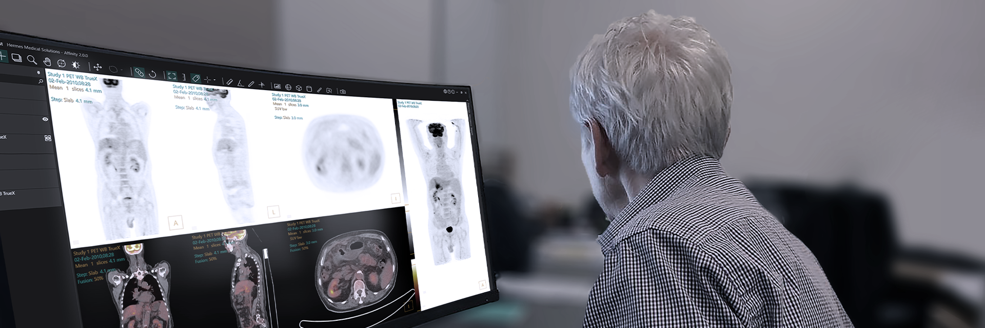
“Hermia is a very user-friendly program, the interface makes a lot of sense and you can customize quite a lot of things in the way you view your images, the overlay in the registration works well and one thing we both find really helpful is the responsiveness of the support when we have a question.”
- Pieter Nienhuis, MD and PhD Candidate
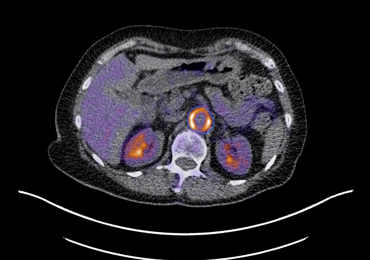
High diffuse 18F-FDG uptake in the abdominal aorta of a patient with large vessel vasculitis.
Using FDG PET to determine inflammation stage and response to treatment
“Diagnosis of large vessel vasculitis is getting better and better with the use of ultrasound for example,” says Pieter. “However, it is very difficult to tell how these patients, that are all started on the same high doses of glucocorticoids, will respond well to that therapy or not. And because of the potential short or long-term side effects of those medications you want to know in advance if they will respond to these. The research may tell if this is possible.” Some research suggests that uptake of FDG may give an indication of how a patient will relapse in the near future. Segmentation of the vessels will be key for the quantification that is necessary to evaluate the FDG uptake, and this is where AI comes in.
“Segmenting vessels is a tedious and long process, and, at the moment, it is not possible to have that in the workflow of a nuclear medicine physician. If it is possible, and it very much looks so, to do the segmentation automatically that would enable the transition of the findings we may have into the clinic. We may find all kind of things but if the clinicians can’t use it there is hardly any point,” notes Pieter.
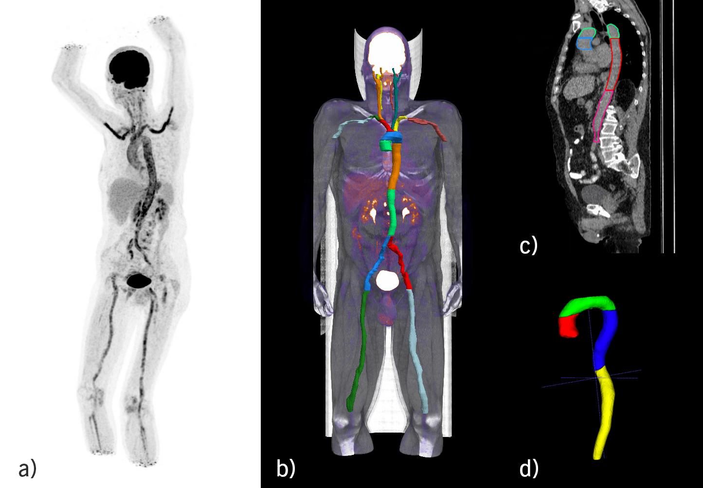
a) Intense 18F-FDG uptake in all large and medium sized vessels in patient with large vessel vasculitis. b) 3D view of manual segmentations of larger and smaller arteries in the Hermia software. c) A low-dose CT with four aortic segments delineated in Hermia: ascending aorta (blue), aortic arch (green), descending aorta (red), and abdominal aorta (magenta). d) 3D rendering of the four aortic segments: ascending aorta (red), aortic arch (green), descending aorta (blue), and abdominal aorta (yellow)
“Hermia is a very user-friendly program, the interface makes a lot of sense and you can customize quite a lot of things in the way you view your images, the overlay in the registration works well and one thing we both find really helpful is the responsiveness of the support when we have a question. We usually get an answer within a day or two. It is really quite amazing,” mentions Pieter.
“The AI model developed by Gijs is trained on low-dose CT datasets, so it won’t matter which tracers will be used. The possibilities are endless of course for any vascular disease,” tells Pieter. “I do believe that once it is working with low-dose CT, the step to go to MR will be easy even if it remains to be seen if contrast differences in PET-MR will matter. Better visibility is expected of small and large vessels on PET-MR (vs low-dose CT) and we are planning to use the same segmentation tools as for PET/CT and expect it to be better - but how much better remains to be seen.” Pieter is currently in Århus Denmark for a research internship focusing on PET-MR.
"To have an independent software tool that can help on that standardization process is very important and your reconstruction software is already one step in the right direction."
- Gijs van Praagh, Biomedical Engineer and PhD candidate
The role of AI in Nuclear Medicine (NM)
“AI will have a supportive and quite big role in NM. It has proven to be very useful in general medical imaging. Due to the better image quality, with sometimes even use of contrast media, it makes sense that the development and implementation started at the radiology department. This makes it quite a lot easier to detect whatever needs to be found in the images compared to a (non-contrast) low- dose CT. However, the impact (of automatic algorithms) in NM might even be bigger because the images are so large,” explains Gijs.
According to Gijs, a major hinder to the implementation of these new tools could be the lack of standardization of PET scans where SUV cut-off values of non-standardized PET images makes things difficult. It is also a general issue for the clinical analysis of those images. “To have an independent software tool that can help on that standardization process is very important and your reconstruction software is already one step in the right direction,” says Gijs.
“At the Hermes Medical Solutions (HMS) user meeting in Rotterdam, it was nice for me as a researcher to be able to show what is possible or not because manufacturers often don’t have all the data that hospitals have. So that first step of getting AI working at hospitals is very important but eventually we want these kinds of tools working in software like Hermia leveraging from the visualizing skills from a software company like HMS.”
Next step to clinical practice
One application from the research that comes closest to clinical practice is differentiating between atherosclerosis and large vessel vasculitis in FDG PET as uptake of the diseases mimic each other. Our aim is to use AI to determine whether there is still active vasculitis to know if a patient needs (more) treatment or whether atherosclerosis is causing the uptake. This could make quite a difference for the treatment strategy of the patient.

 a) Intense 18F-FDG uptake in all large and medium sized vessels in patient with large vessel vasculitis.