Implementation of Hermia Dosimetry Software: Initial Clinical Experience from Ankara University
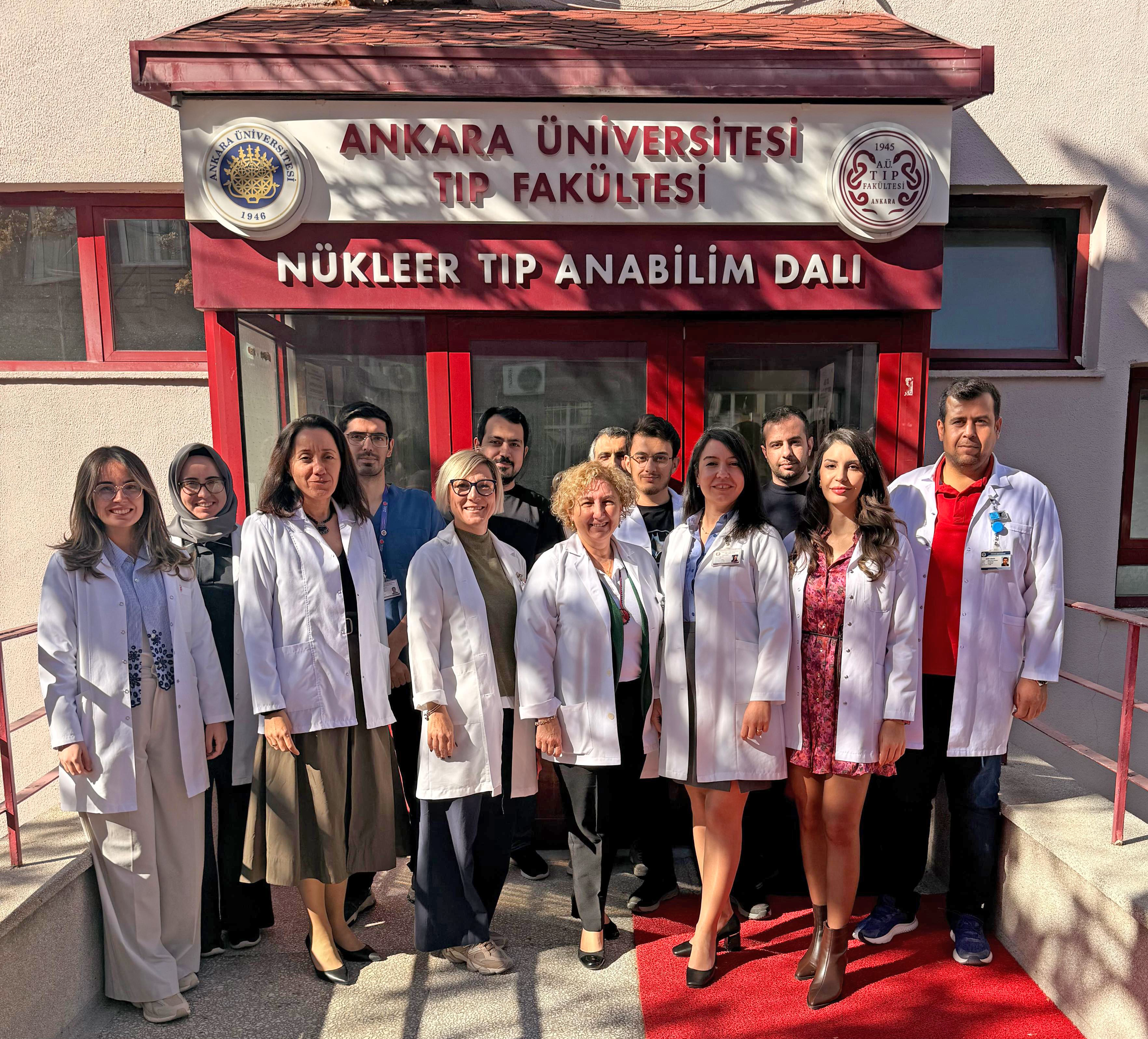
As the centre where the Hermia software was first installed in Turkey—a country at the forefront of patient volume and academic research in radioembolisation—we aimed to share our initial experience and perspectives with the medical physics community. The Department of Nuclear Medicine at Ankara University is among the country’s leading centres for radionuclide therapies, including transarterial radioembolisation (TARE), I-131, Lu-177 DOTATATE, and Lu-177 PSMA therapies. Approximately one hundred radioembolisation procedures are performed annually at our centre, addressing both primary and metastatic liver tumours. The dosimetry software our centre utilises is Hermia SIRT (Selective Internal Radiation Therapy). Hermia SIRT follows the EANM guidelines and uses computer-assisted liver, lung, and lesion segmentation, live dose feedback and allows planning and verification of multiple injections.
The local context
Over the past decade, the field of radioembolization has evolved substantially, particularly with the growing emphasis on personalized dosimetry to maximize therapeutic benefit while minimizing hepatic and systemic toxicity. This shift from empirical to quantitative treatment planning has created a demand for robust, validated, and user-friendly dosimetry platforms suitable for routine clinical practice. With this in mind, our department has long sought a reliable solution that combines simplicity with quantitative accuracy. Following positive interactions with Hermes Medical Solutions during both national and EANM congresses, we decided to implement the Hermia dosimetry software in our clinical workflow. After a concise online training session, the system was rapidly integrated into daily practice.
Initial experience
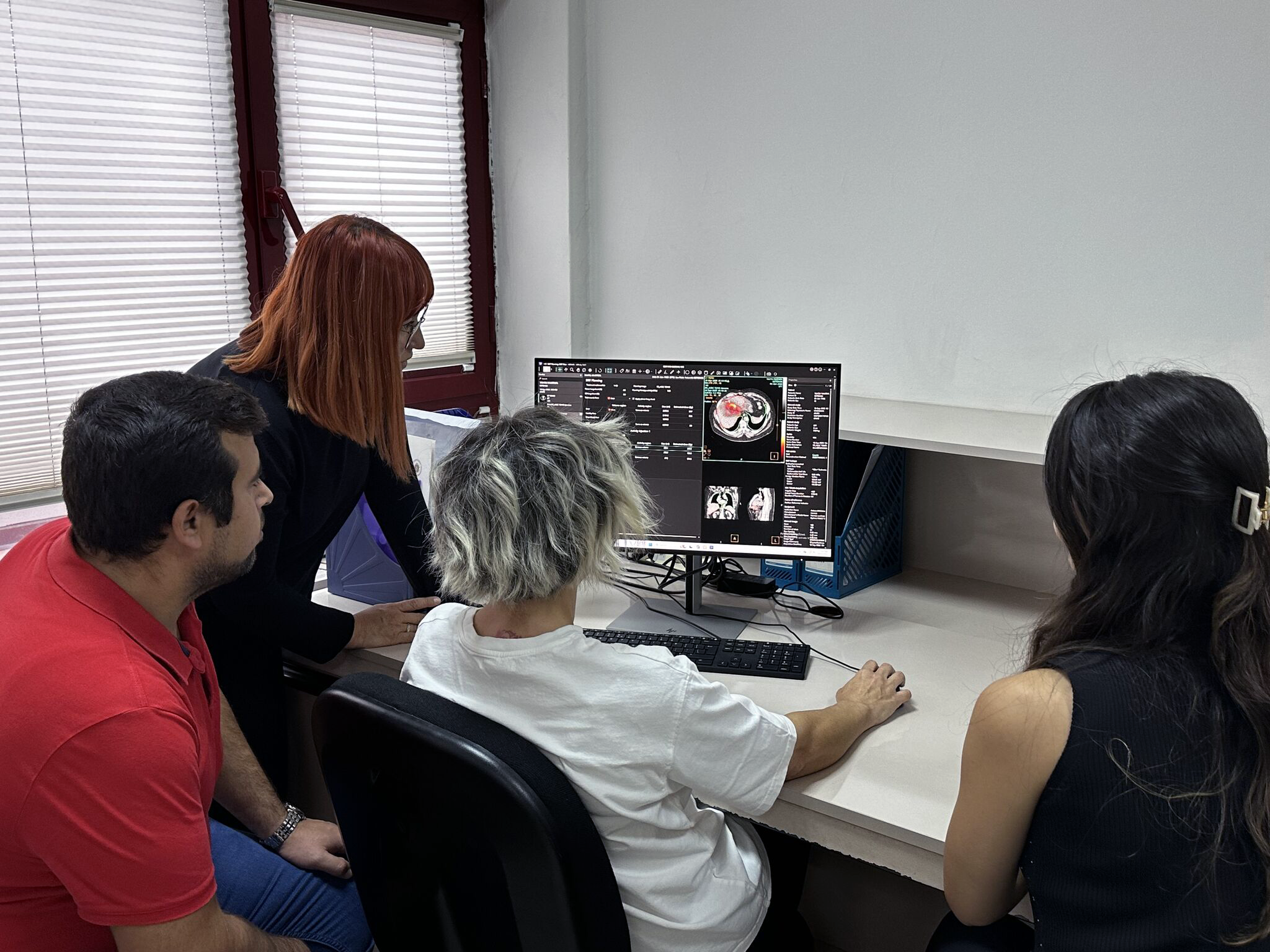
Our early experience revealed that Hermia’s user interface provides remarkable workflow efficiency. The automatic segmentation of organs on CT datasets significantly reduces manual processing time. The one-click segmentation of perfused liver volumes and tumours is a particularly valuable feature, especially in centres with a high procedural workload. Additionally, treatment verification can be swiftly performed using post-therapy Y-90 PET/CT images without the need for re-segmentation, ensuring consistency between pre- and post-treatment dosimetry. These features collectively enable fast, reproducible, and clinically practical personalized dosimetry, aligning well with the increasing focus on patient-specific treatment optimization.
From a research perspective, the workstation has streamlined our post-treatment dosimetric analyses, facilitating retrospective and prospective investigations with greater ease. The ability to export voxel-based dose maps and quantitative data directly from the Hermia platform supports reliable dose–response and toxicity correlation studies. Such improvements in data handling are expected to accelerate our scientific output and enhance the precision of dosimetry-based clinical decision-making. We anticipate that these capabilities will strengthen multidisciplinary collaborations among nuclear medicine, radiology, and medical physics teams within our institution.
 Next steps
Encouraged by this initial success, our next step will be to incorporate the voxel-based dosimetry module into our clinical and research protocols. This extension will enable a more comprehensive assessment of spatial dose heterogeneity, especially in liver parenchyma and critical structures, thereby improving our capacity to conduct detailed dose-effect modelling. The integration of voxel-level dosimetry will also contribute to our ongoing clinical trials investigating dose thresholds and radiobiological response in novel radionuclide therapies.
Summary
In summary, the implementation of the Hermia dosimetry solution from Hermes Medical Solutions at our centre has proven both feasible and beneficial for routine practice and research. Its intuitive interface, automation capabilities, and compatibility with multimodal imaging workflows have greatly improved efficiency and reproducibility. The software’s seamless integration into TARE procedures exemplifies the potential of advanced dosimetry tools to promote the widespread adoption of quantitative, patient-centered treatment planning in nuclear medicine.
 Biography